

What Is an Oral Biopsy?

When a Finding Needs to Be Accurately Identified.

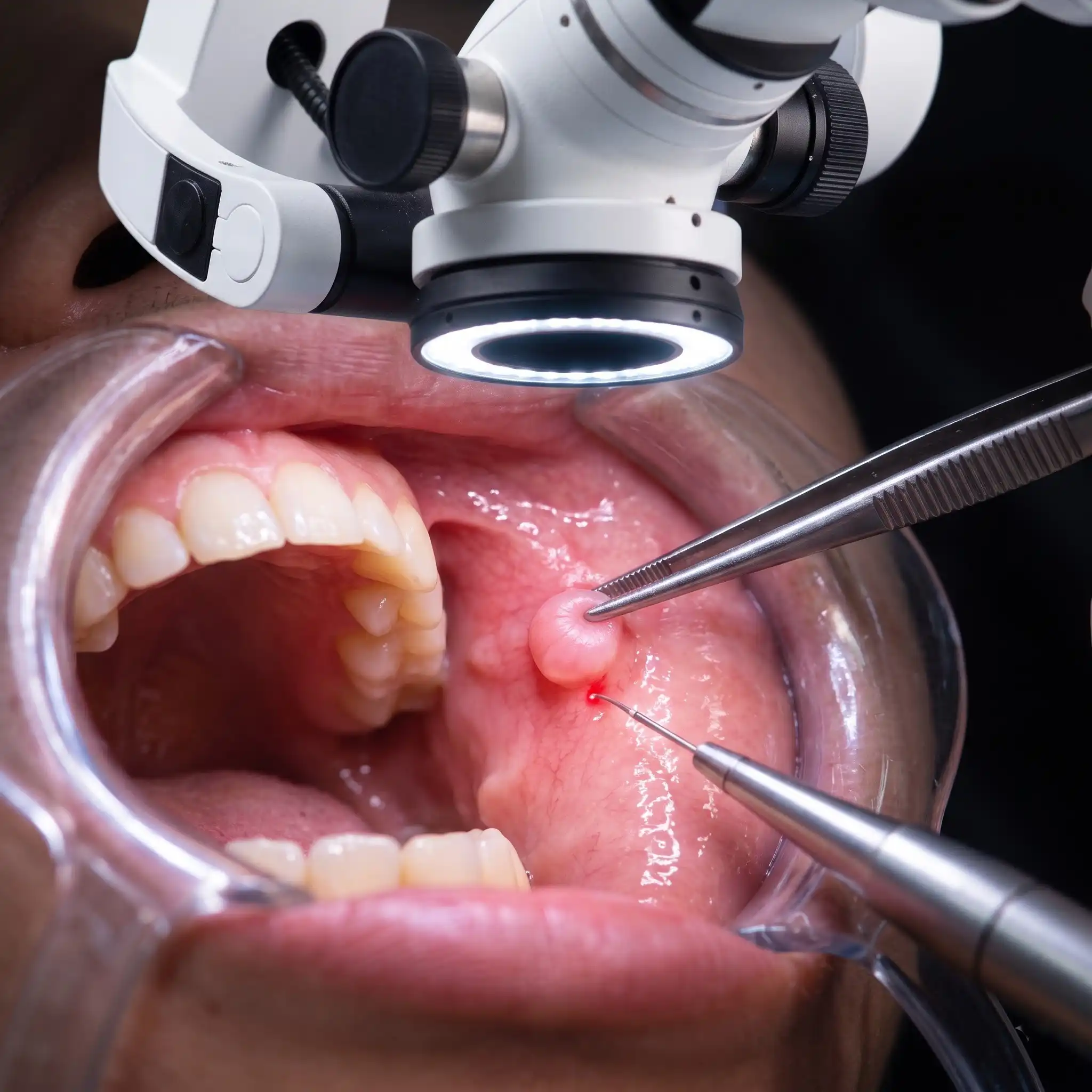
An oral biopsy is a procedure in which a small sample of tissue is removed from an abnormal area in the mouth, a lesion, ulcer, white or red patch, or growth that has not resolved on its own, for detailed laboratory examination. The biopsy allows the tissue to be assessed under a microscope, providing an accurate diagnosis of what the finding represents.

Oral biopsies are not a cause for alarm in themselves; they are a diagnostic procedure used to establish precisely what a suspicious area is so that the appropriate management can be determined. Many biopsied lesions are benign. The value of the biopsy is in establishing this with certainty, rather than monitoring indefinitely without a clear answer.

At Halo 32, oral biopsies are performed using advanced soft tissue laser technology, providing exceptional precision, minimal bleeding, and faster healing than conventional surgical biopsy.

When Is a Biopsy Recommended?

When an Area in the Mouth Requires a Definitive Answer.

Your dentist may recommend a biopsy if an area in the mouth has not resolved within two to three weeks without an identifiable cause, or if its appearance warrants closer examination. Common indications include:

  • A persistent ulcer or sore that has not healed after two to three weeks
  • A white patch (leukoplakia), red patch (erythroplakia), or mixed red-and-white lesion
  • A lump, thickening, or growth in the soft tissue of the mouth
  • A lesion with irregular borders, uneven surface, or unusual colour
  • A previously identified lesion that has changed in size, shape, or appearance
  • Suspicious tissue identified during a routine oral cancer screening

The majority of biopsied oral lesions are benign, but identification requires tissue analysis, not observation alone. Early identification of any concerning finding is always in the patient’s best interest.

THE HALO 32 PROCESS

Your Biopsy Appointment at Halo 32

01

Clinical Assessment

The area is examined and documented before biopsy is performed. The location, size, appearance, and clinical characteristics of the lesion are recorded. The decision to biopsy, and the type of biopsy most appropriate, is made based on this assessment.

02

Local Anaesthesia

The area is fully anaesthetised before the biopsy begins. You will feel no pain during the procedure, only mild pressure at most.

03

Laser Biopsy

The soft tissue laser removes a precise sample of the abnormal tissue with exceptional accuracy, sealing the tissue simultaneously. Bleeding is minimal. In most cases, no sutures are required. The procedure is completed quickly, typically within fifteen to thirty minutes, and the sample is sent to a laboratory for histological analysis.

04

Results & Next Steps

Laboratory results are typically available within one to two weeks. Your dentist will contact you to discuss the findings and, where relevant, recommend the most appropriate next steps, whether that is monitoring, further treatment, or referral. No finding is communicated without a full explanation of what it means and what the options are.

What a Laser Biopsy Does for You

Early Identification Where It Matters

Early identification of any oral tissue change, whether benign, pre-malignant, or malignant, allows for the most straightforward and most effective management. A biopsy performed promptly on a concerning lesion is always preferable to delayed investigation.

Completed in One Appointment

The biopsy itself, assessment, anaesthesia, laser excision, and dressing are completed in a single appointment. You leave the same day with the sample submitted for analysis and a clear timeline for results.

An Accurate Diagnosis

A biopsy provides a definitive tissue diagnosis, something that clinical observation, imaging, and examination cannot. For any lesion with uncertain identity, a biopsy replaces uncertainty with a clear answer.

Laser Precision, Less Trauma

The soft tissue laser removes the biopsy sample with a level of precision that minimises disruption to the surrounding healthy tissue. Less trauma means less bleeding during the procedure, less post-operative discomfort, and faster healing.

What to Expect After Your Biopsy

A Minor Procedure. A Clear Answer.

Recovery from a laser biopsy is typically straightforward. The laser sealing during the procedure reduces post-operative bleeding and discomfort significantly compared to conventional surgical biopsy.

  • Day 1–3: Mild tenderness in the biopsied area is normal. Standard pain relief is usually sufficient.
  • Diet: Soft foods and cool or room-temperature drinks for the first few days. Avoid anything that might irritate the healing area.
  • Oral Hygiene: Continue brushing gently around the area. Specific aftercare guidance will be provided before you leave.
  • Results: Laboratory results are typically available within one to two weeks. Your dentist will contact you to discuss findings and next steps.

Why Halo 32 for Oral Biopsy

Laser Technology as Standard

Oral biopsies at Halo 32 are performed with soft tissue laser technology, not conventional scalpel and suture technique. The precision and reduced trauma of laser biopsy are particularly important in oral soft tissue, where the quality of the sample and the minimisation of surrounding tissue damage both matter.

Results Communicated With Care

Biopsy results are discussed with you directly, with full explanation of what the findings mean, what options are available, and what the recommended next steps are. Nothing is communicated without context, and no finding is left unexplained.

Part of a Comprehensive Oral Health Assessment

At Halo 32, oral cancer screening is part of every routine check-up. Where a biopsy is recommended, it follows from a systematic assessment, not an incidental finding. Early identification is built into the standard of care here.
FAQs

Does a biopsy mean I have cancer?

No. A biopsy is a diagnostic procedure; its purpose is to identify what a tissue finding is, not to confirm a specific diagnosis. The majority of biopsied oral lesions are benign. A biopsy is recommended whenever a finding cannot be confidently identified by clinical assessment alone, which applies to many entirely harmless conditions. Early investigation is always preferable to delayed certainty.

Laboratory histological analysis typically takes one to two weeks. Your dentist will contact you directly when results are available to discuss the findings and any recommended next steps.

Small oral biopsies heal with minimal visible change to the tissue. Laser biopsy produces less scarring than conventional surgical technique because the laser seals and minimises trauma to surrounding tissue during the procedure. Healing is typically clean and complete within two to three weeks.

The procedure is performed under local anaesthesia; you will feel no pain during the biopsy. Post-operative tenderness for one to three days is normal and manageable with standard pain relief.

A Clear Answer, Obtained With Precision and Care.

Book your consultation at Halo 32 in Kharadi, Pune.

Reserve Your Appointment

Every great smile begins with a conversation. Book your consultation at Halo 32 and take the next step towards personalised dental care.

Call us Chat on WhatsApp