
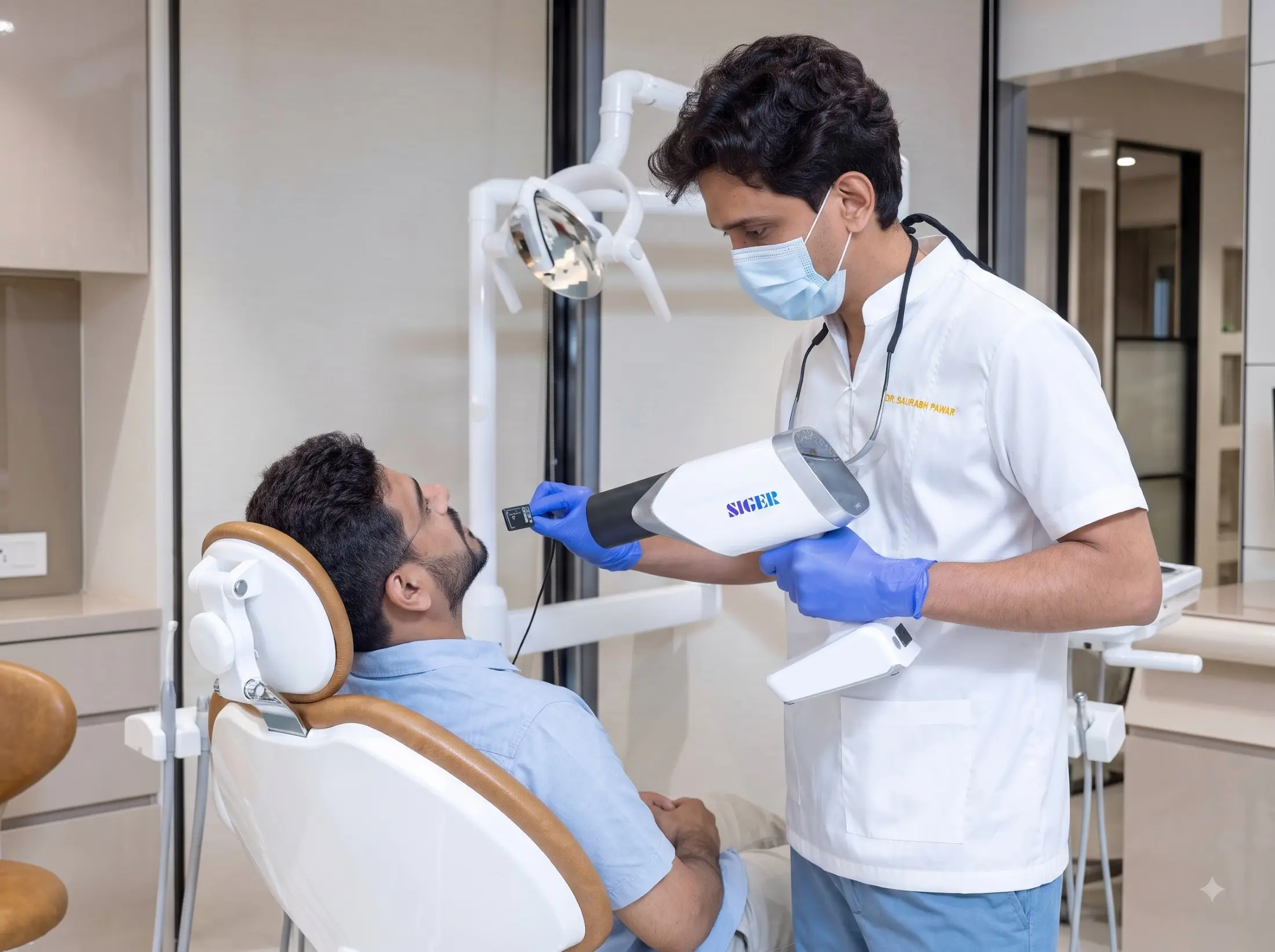

What Are Dental X-Rays?

The most consequential dental problems – decay between teeth, bone loss, root fractures, infections forming in silence – are invisible to the naked eye until they have already caused significant damage. Digital dental X-rays surface all of it. Early, accurately, and with a fraction of the radiation of conventional X-ray systems.
At Halo 32, RVG digital X-ray technology produces high-definition images instantly, displayed on screen for you to see alongside your dentist, with findings explained clearly as they are reviewed. There are no film delays, no guesswork, and no findings communicated without full explanation.

When Are Dental X-Rays Needed?

X-rays are taken when a clinical question cannot be answered by examination alone. You may need dental X-rays if:

  • You are a new patient and a baseline record of your teeth and jaw is required
  • You have toothache, sensitivity, or swelling and the cause is not visible
  • Your dentist suspects decay between teeth or beneath an existing filling or crown
  • Your gums show signs of disease and bone levels need to be assessed
  • You have had trauma to the teeth or jaw
  • You are being assessed for root canal treatment, wisdom tooth removal, implants, or orthodontics

How often you need X-rays depends on your individual oral health, not a fixed schedule. Patients with healthy teeth and gums need them far less frequently than those with active decay or gum disease, and at Halo 32 the interval is set accordingly.

TYPES OF X-RAYS USED AT HALO 32

The Right Image for the Right Clinical Question.

01

Bitewing X-rays

Show the crowns of the upper and lower back teeth simultaneously; the most effective tool for detecting decay between teeth and monitoring existing restorations.

02

Periapical X-rays

Show the full length of one or two teeth, roots, surrounding bone, and any pathology at the root tip. Used to investigate specific symptoms or findings.

03

Panoramic X-rays (OPG)

A single image of the entire mouth, all teeth, both jaws, the temporomandibular joints, and the sinuses. Used for treatment planning, orthodontic assessment, and implant planning.

04

CBCT (Cone Beam CT)

Three-dimensional imaging of the jaw, bone, and surrounding structures. Used for implant planning, complex surgical cases, and detailed assessment of anatomical structures that two-dimensional imaging cannot fully capture.

What Dental X-Rays Reveal

Problems Beneath Restorations

Decay can develop under existing fillings and crowns where it cannot be seen. X-rays show the condition of the tooth beneath, so failing restorations are replaced before damage becomes extensive.

Infection at the Root

An abscess forming at the tip of a root can develop for months without symptoms. X-rays reveal it, allowing root canal treatment before the infection spreads or the tooth is lost.

Bone Loss From Gum Disease

Gum disease destroys the bone that supports the teeth, often without pain. X-rays measure bone levels accurately, so loss is identified and treated before teeth become loose.

Decay Between Teeth

The surfaces where teeth touch cannot be seen during an examination, yet they are among the most common sites of decay. Bitewing X-rays detect it early, while a small filling is still enough.

Impacted and Developing Teeth

X-rays show teeth that have not yet erupted, including impacted wisdom teeth and developing teeth in children, allowing problems with position or eruption to be planned for early.

Precise Treatment Planning

Root canals, extractions, implants, and orthodontics all depend on knowing the exact anatomy of the teeth and jaw. X-rays provide the measurements every treatment plan is built on.

Are Dental X-Rays Safe?

Safe. Targeted. Used Only When Indicated.

Digital X-rays at Halo 32 use RVG technology, which delivers a fraction of the radiation of conventional film X-rays, significantly less than what you are exposed to on a short flight. X-rays are taken only where clinically indicated, not as a routine at every visit. Every image is taken with a specific clinical purpose, and findings are reviewed with you directly before any treatment discussion.

Why Halo 32 for Dental X-Rays

Instant, High-Definition Imaging

RVG digital sensors produce detailed images within seconds. There is no waiting for film to develop and no repeat exposures from unclear results; images can be enlarged and enhanced on screen to reveal detail film cannot show.

Reviewed With You, On Screen

Every X-ray is displayed on screen and walked through with you. You see the same image your dentist sees, with each finding explained in plain terms before any treatment is discussed.

Imaging That Informs Treatment

At Halo 32, X-rays are not a separate step. Images feed directly into treatment planning for root canals, implants, orthodontics, and restorations, so every recommendation is based on what the imaging actually shows.

FAQs

How often do I need dental X-rays?
For most patients in good dental health, X-rays are needed every one to two years. Patients with a history of decay, gum disease, or active treatment may require more frequent imaging. We take X-rays where they are clinically justified, not on a fixed schedule regardless of need.
Routine dental X-rays are generally avoided during pregnancy as a precautionary measure, particularly in the first trimester. Where an X-ray is clinically necessary during pregnancy, appropriate shielding is used. Your dentist will discuss this with you if relevant.
Yes, always. At Halo 32, every X-ray is reviewed on screen with you directly. We walk through what each image shows, explain any findings in plain language, and answer any questions before treatment is discussed.

A Clear Picture of Your Oral Health.

Book your appointment at Halo 32 in Kharadi, Pune.

Reserve Your Appointment

Every great smile begins with a conversation. Book your consultation at Halo 32 and take the next step towards personalised dental care.

Call us Chat on WhatsApp